Magnetic Resonance Imaging (MRI)
About the MRI - Basic Description
MRI is a technique for viewing the brain's structure and functions. Two main forms exist: structural MRI provide detailed pictures of the brain's shape and size. Functional MRI allows researchers to visualize and map the parts of the brain used to perform everyday tasks, such as reading and calculation. Both structural and functional MRI are used for our studies in CIBSR.
The MRI machine is, in essence, a big magnet. As you lie in its magnetic field, invisible radio waves are released around you. This will result in harmless radio waves bouncing off the different substances that make up your brain. These radio waves are then detected by a computer, which transforms the data into images of the brain's structure and activity.
In functional MRI (fMRI), as you lie in the MRI machine, you are given simple tasks, like math addition or subtraction; the MRI then maps what parts of the brain are most active during those tasks compared with activity while the brain is at rest. The areas of most activity in individuals with a healthy brain are then compared to individuals with a disorder like fragile X syndrome. This allows researchers to understand how the brain functions; this information is used together with the data from the structural MRI data to piece together a comprehensive picture of brain structure and function that fit in an overall picture of the disorders we study. Our structural and functional MRI studies also allow us to understand how the healthy brain works.
About the MRI - Detailed Description
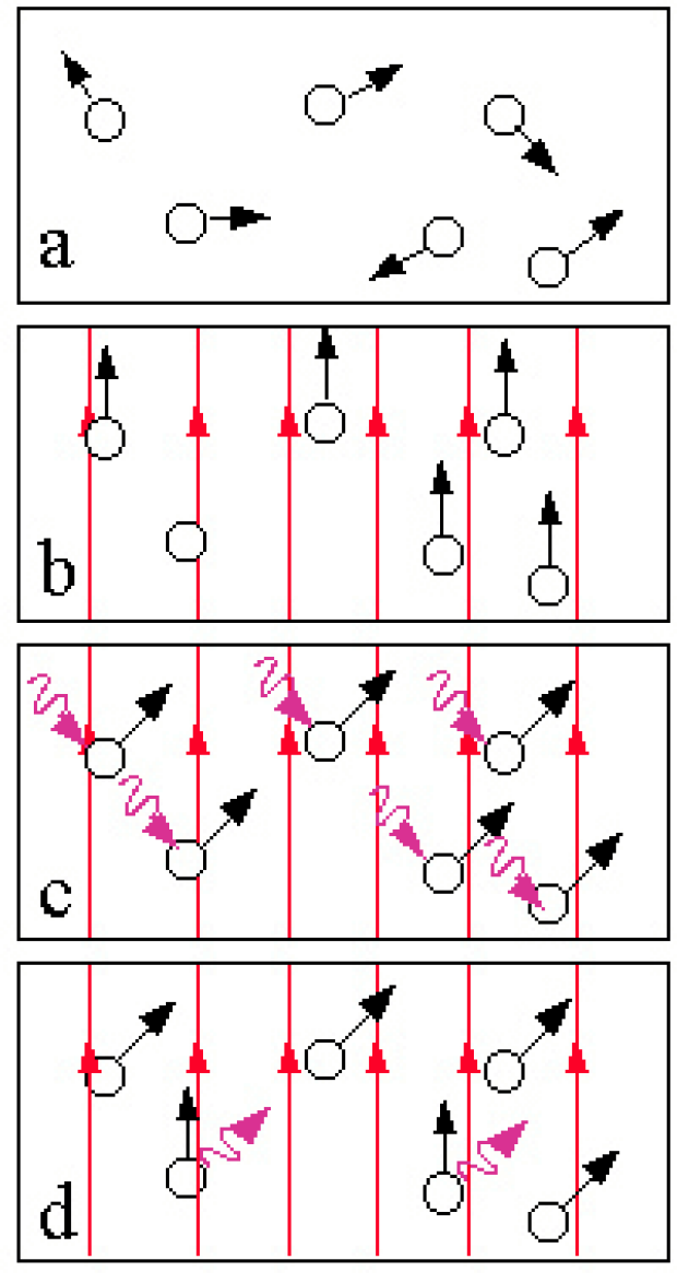
Magnetic resonance imaging (MRI) generates cross-sectional images of the human body by using nuclear magnetic resonance (NMR). The process begins with positioning the imaged body (a) in a strong, uniform magnetic field, which polarizes the nuclear magnetic moments of water protons by forcing their spins into one of two possible orientations (b). Then an appropriately polarized radio-frequency field, applied at resonant frequency, forces spin transitions between orientations (c). Those transitions create a signal (d) (which is an NMR phenomenon) that can be detected by a receiving coil.
An MRI scanner applies the radio-frequency field as finely crafted pulses, which excite only protons whose resonant frequencies fall within a fairly narrow range. Applying magnetic-field gradients during the radio-frequency pulse creates resonant conditions for only the protons that are located in a thin, predetermined slice of the body. Orientation and thickness of this slice can be selected arbitrarily in the imaged body. The NMR signal encodes positional information across the slice by using a method known as the ``spin warp,'' and a two-dimensional Fourier Transform extracts that positional information. The process creates a data matrix in which each element represents an NMR signal from a single, localized volume element, or voxel, within the imaged slice. A two-dimensional display of this matrix's contents creates a human-readable image of the selected slice. Each image element, or pixel, represents the NMR signal strength that was recorded for its corresponding voxel.
An MRI image provides unmatched soft-tissue contrast. When compared with other medical-imaging techniques, MRI provides several significant advantages: noninvasiveness, safety (because it uses non-ionizing radiation), and superb soft-tissue contrast, generated by an NMR signal's sensitivity to tissue morphology and pathology.
For more information: http://biac.stanford.edu