Near-Infrared Spectroscopy (NIRS)
About NIRS - Basic Description
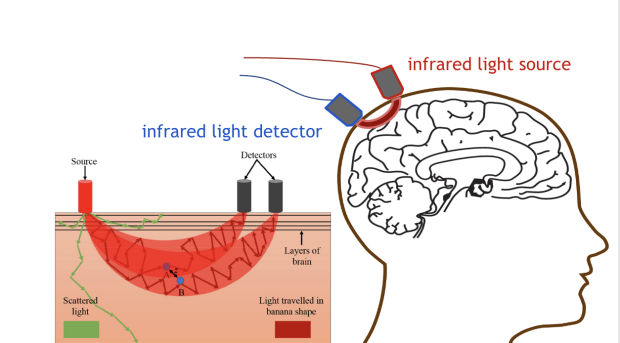
Functional near-infrared spectroscopy (fNIRS) is a specialized research optical imaging technique that uses near-infrared light to examine the function of the living brain. Research participants can be standing, sitting, lying down, or even engaged in active behaviors (walking, driving, exercising) while receiving an fNIRS scan. Small probes are placed on the top of the head using a cap. There is no sensation of any kind from the infrared light, and it is not harmful. An actual fNIRS scan typically takes less than one hour (depends on tasks). A participant might be asked to engage in a variety of activities during an fNIRS scan. Some activities might involve passively listening to sounds or seeing flashes, or performing learning, memory or other behavioral tasks. Participants might also be asked to play certain games or engage in activities on the computer or with an experimenter during the scan.
About NIRS - Detailed Description